简体中文

AC-4-细颗粒型

同义词 细颗粒 fine granular
描述
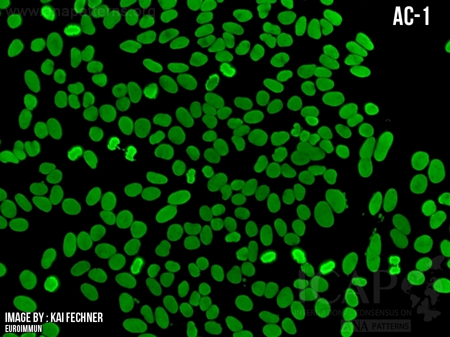
细胞核中散布细小的颗粒样荧光,核仁通常无荧光。有丝分裂期细胞(中期、后期和末期)染色质无荧光,染色质周围有荧光。
Fine tiny speckles dispersed throughout the nucleoplasm. The nucleoli are typically not stained. The chromatin mass of mitotic cells (metaphase, anaphase, and telophase) is, in contrast to the cytoplasm surrounding the chromatin mass, not stained.
抗原相关性 SS-B/La, Mi-2, TIF1γ, TIF1β, Ku, TROVE2/Ro60(此前被称为SS-A/Ro60 [1] ,并经常与其他抗体一起出现) 注释#1.抗SS-A/Ro60抗体在HEp-2000载片(IMMUNO CONCEPTS)上的荧光染色与HEp-2载片不同。 SS-B/La, Mi-2, TIF1γ, TIF1β, Ku, TROVE2/Ro60 (formerly known as SS-A/Ro60 [1] and often in combination with antibodies to other antigens).Note #1. Anti-SS-A/Ro60 staining on HEp-2000 slides (IMMUNO CONCEPTS) is different from other HEp-2 slides.
  • 临床相关性

    一级信息

    关于临床相关性和缩写列表

    Clinical Relevance

    First level information

    About Clinical Relevance & List of Abbreviations
  • ▶这是实验室HEp-2 IFA常规检测中最常见的核型,也是在荧光阳性但无临床症状或无系统性自身免疫性疾病的患者中最常观测到的核型之一。
    ▶在不同的系统性自身免疫性疾病(SARD)中均有不同程度的表现,尤其是原发性干燥综合征(SjD),系统性红斑狼疮(SLE),亚急性皮肤型红斑狼疮(SCLE),新生儿红斑狼疮(NLE),特发性炎症性肌病(IIM),系统性硬化症(SSc)以及SSc-IIM重叠综合征。
    ▶如果临床怀疑SjD、SLE、SCLE、NLE或先天性心脏传导阻滞等情况,建议进行针对抗TROVE2/Ro60抗体的检测。
    ▶抗TROVE2/Ro60抗体是干燥综合征诊断标准的一部分(注:这些标准以及某些商业免疫检测方法并未区分TROVE2/Ro60与TRIM21/Ro52)[2]
    ▶如果临床怀疑存在IIM(或程度较轻的SSc-IIM和SLE-IIM)的重叠综合征,则建议进一步检测抗Mi-2、TIF1γ和Ku自身抗体;这些抗原通常包含在疾病特异性免疫检测中(如炎症性肌病谱) [3,4]。
    ▶抗Mi-2和抗TIF1γ抗体与皮肌炎(DM)有关,在患有皮肌炎的患者中,抗TIF1γ抗体虽然在总体的AC-4核型中较为罕见,但与年龄>40岁的患者的恶性肿瘤具有很强的关联性[3, 5, 6]。
    ▶抗Ku抗体与SSc-IIM和SLE-IIM有关,且具有独特的临床特征[4, 7]
    ▶由于AC-4和AC-31核型极为相似,但却具有不同的临床意义,建议区分两种核型[8]。
    在某些样本中,抗TROVE2/Ro60抗体和IIM特异性自身抗体可能无法在HEp-2免疫荧光筛查中被检测到[9]。
    对于抗TRIM21/Ro52抗体(以前被称为SS-A/Ro52[1])是否会产生AC-4核型尚不明确。而表现出AC-4核型的学期可能会含有抗TRIM21/Ro52抗体,单特异性抗TRIM21/Ro52抗体在HEp-2免疫荧光检测中并不总是显示出染色现象[10]。
    This is the most frequent pattern in general laboratory HEp-2 IFA routine testing and one of the most frequent patterns observed in HEp-2 IFA-positive asymptomatic individuals and patients with no systemic autoimmune disease
    ▶Present to a varying degree in distinct systemic autoimmune rheumatic diseases (SARD), in particular primary Sjögren’s disease (SjD), systemic lupus erythematosus (SLE), subacute cutaneous lupus erythematosus (SCLE), neonatal lupus erythematosus (NLE), idiopathic inflammatory myopathies (IIM), systemic sclerosis (SSc), and SSc-IIM overlap syndrome
    If SjD, SLE, SCLE, NLE, or congenital heart block is clinically suspected, it is recommended to perform follow-up tests for anti-TROVE2/Ro60
    Autoantibodies to TROVE2/Ro60 are part of the classification criteria for SjD (note: these criteria and some commercial immunoassays do not distinguish between TROVE2/Ro60 and TRIM21/Ro52) [2]
    If IIM, or to a lesser extent SSc-IIM and SLE-IIM overlap syndromes, is clinically suspected, follow-up tests for detecting autoantibodies to Mi-2, TIF1γ, and Ku is recommended; these antigens are typically included in disease specific immunoassays (i.e., inflammatory myopathy profile) [3, 4]
    Autoantibodies to Mi-2 and TIF1γ are associated with dermatomyositis (DM); autoantibodies to TIF1γ in patients with DM, although rare in the overall AC-4 pattern, is strongly associated with malignancy in patients >40 years old [3, 5, 6]
    Autoantibodies to Ku are associated with SSc-IIM and SLE-IIM overlap syndromes with a unique phenotype [4, 7]
    For optimal clinical relevance the AC-4 pattern should be distinguished from the AC-31 pattern, as these are closely resembling patterns that have potentially different clinical significance [8]

    Anti-TROVE2/Ro60 and IIM-specific autoantibodies may be undetected in HEp-2 IFA-screening in some substrates [9]

    There is often uncertainty regarding whether anti-TRIM21/Ro52 antibodies (formerly SS-A/Ro52 [1]) produce an AC-4 pattern. While sera exhibiting an AC-4 pattern may contain anti-TRIM21/Ro52 antibodies, monospecific anti-TRIM21/Ro52 sera do not consistently show staining in HEp-2 IFA [10]

  • 二级信息
    Second level information
  • None
  • 参考文献
  • 1.Choi M, Andrade LEC, Chan EKL, Fritzler MJ, Ben-Chetrit E, et al. Autoantibody nomenclature harmonization: Ro, SSA, TRIM21 and TROVE2. in preparation. 2025
    2.Shiboski CH, Shiboski SC, Seror R, Criswell LA, Labetoulle M, Lietman TM, Rasmussen A, Scofield H, et al. 2016 American College of Rheumatology/European League Against Rheumatism classification criteria for primary Sjogren's syndrome: A consensus and data-driven methodology involving three international patient cohorts. Ann Rheum Dis. 2017;76:9-16
    3.Choi MY, Satoh M, Fritzler MJ. Update on autoantibodies and related biomarkers in autoimmune inflammatory myopathies. Curr Opin Rheumatol. 2023;35:383-94
    4.Betteridge Z, McHugh N. Myositis-specific autoantibodies: an important tool to support diagnosis of myositis. J Intern Med. 2016;280:8-23
    5.Trallero-Araguas E, Rodrigo-Pendas JA, Selva-O'Callaghan A, Martinez-Gomez X, Bosch X, Labrador-Horrillo M, Grau-Junyent JM, Vilardell-Tarres M. Usefulness of anti-p155 autoantibody for diagnosing cancer-associated dermatomyositis: a systematic review and meta-analysis. Arthritis Rheum. 2012;64:523-32
    6.Wang G, McHugh NJ. An update on myositis autoantibodies and insights into pathogenesis. Clin Exp Rheumatol. 2025;43:364-71
    7.Holzer MT, Uruha A, Roos A, Hentschel A, Schanzer A, Weis J, Claeys KG, Schoser B, et al. Anti-Ku + myositis: an acquired inflammatory protein-aggregate myopathy. Acta Neuropathol. 2024;148:6
    8.Andrade LEC, Klotz W, Herold M, Musset L, Damoiseaux J, Infantino M, Carballo OG, Choi M, et al. Reflecting on a decade of the international consensus on ANA patterns (ICAP): Accomplishments and challenges from the perspective of the 7th ICAP workshop. Autoimmun Rev. 2024;23:103608
    9.Bossuyt X, Frans J, Hendrickx A, Godefridis G, Westhovens R, Marien G. Detection of anti-SSA antibodies by indirect immunofluorescence. Clin Chem. 2004;50:2361-9.
    10.Chan EKL. Anti-Ro52 Autoantibody Is Common in Systemic Autoimmune Rheumatic Diseases and Correlating with Worse Outcome when Associated with interstitial lung disease in Systemic Sclerosis and Autoimmune Myositis. Clin Rev Allergy Immunol. 2022;63:178-93

FAQ