| 简体中文 | ||
| 同义词 | MSA-3, NSp-II | MSA-3, NSp-II |
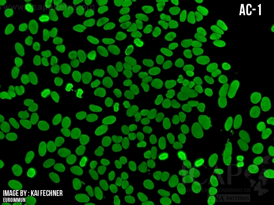
| 描述 | 在不同的细胞周期中,核斑点染色强度有显著差异,G2期染色最强,G1期最弱或无染色。着丝粒体仅在前中期和分裂中期细胞内呈阳性,显示出多个排列整齐但小而微弱的斑点。在前中期细胞中,细胞核膜常常呈现出较弱的染色现象。某些细胞在分裂后期和末期会呈现出子细胞分裂中区环(即中间体和茎体)的强烈染色。分裂期细胞的细胞质呈现弥漫性染色。 |
Nuclear speckled pattern with striking variability in intensity with the strongest staining in G2 and weakest/negative staining in G1 of the cell cycle. The centromeres are positive only in prometaphase and metaphase cells, revealing multiple aligned small and faint dots. Prometaphase cells frequently show a weak staining of the nuclear envelope. During anaphase and telophase, some sera demonstrate intense staining of the mid-body and stem body between the daughter cells. The surrounding cytoplasm of the mitotic cells is diffusely stained. |
| 抗原相关性 | CENP-F (着丝点蛋白F) | CENP-F (centromere protein F) |
-
▶大多数表现出AC-14核型的血清来自患有多种肿瘤病症(乳腺癌,肺癌,结肠癌,淋巴瘤,卵巢癌,脑癌)的患者;矛盾的是,患有这些恶性肿瘤的患者中出现AC-14核型的概率很低 (1-3)。▶AC-14核型也可见于炎症性疾病(克罗恩病、自身免疫性肝病、干燥综合征、移植物抗宿主病);目前关于临床相关性的信息主要基于病例报告和一系列病例结果。▶目前针对抗CENP-F抗体的特异性免疫检测尚无商品化试剂(3-7)。▶The majority of sera exhibiting the AC-14 pattern are from patients with a diversity of neoplastic conditions (breast, lung, colon, lymphoma, ovary, brain); the frequency of the AC-14 pattern in patient cohorts with these malignancies is paradoxically low (1-3).▶The AC-14 pattern is also seen in inflammatory conditions (Crohn’s disease, autoimmune liver disease, Sjögren’s disease, graft-versus-host disease); current information on clinical associations is based mainly on case reports and series of cases.▶Specific immunoassays for CENP-F autoantibody are currently not commercially available (3-7).
-
二级信息Second level information
-
▶其他与AC-14不同的核多形性型应被归类为未分类模式(AC-XX),并附上适当的描述。▶Other nuclear pleomorphic patterns that are distinct from AC-14 should be reported as unclassified patterns (AC-XX) with an appropriate description.
-
参考文献
-
1.Le Joncour A, Charuel JL, Choquet S, Spano JP, Corvol JC, Fautrel B, et al. Diseases associated with anti-centromere protein F (anti-CENP-F) antibodies: Insights from a large cohort study. Joint bone spine. 2025;92(5):1058852.Fritzler MJ, Rattner JB, Luft LM, Edworthy SM, Casiano CA, Peebles C, et al. Historical perspectives on the discovery and elucidation of autoantibodies to centromere proteins (CENP) and the emerging importance of antibodies to CENP-F. Autoimmunity reviews. 2011;10(4):194-2003.Welner S, Trier NH, Frisch M, Locht H, Hansen PR, Houen G. Correlation between centromere protein-F autoantibodies and cancer analyzed by enzyme-linked immunosorbent assay. Molecular cancer. 2013;12(1):954.Casiano CA, Landberg G, Ochs RL, Tan EM. Autoantibodies to a novel cell cycle-regulated protein that accumulates in the nuclear matrix during S phase and is localized in the kinetochores and spindle midzone during mitosis. Journal of cell science. 1993;106 ( Pt 4)(Pt 4):1045-565.Casiano CA, Humbel RL, Peebles C, Covini G, Tan EM. Autoimmunity to the cell cycle-dependent centromere protein p330d/CENP-F in disorders associated with cell proliferation. Journal of autoimmunity. 1995;8(4):575-866.Rattner JB, Rees J, Whitehead CM, Casiano CA, Tan EM, Humbel RL, et al. High frequency of neoplasia in patients with autoantibodies to centromere protein CENP-F. Clinical and investigative medicine Medecine clinique et experimentale. 1997;20(5):308-197.Bencimon C, Salles G, Moreira A, Guyomard S, Coiffier B, Bienvenu J, et al. Prevalence of anticentromere F protein autoantibodies in 347 patients with non-Hodgkin's lymphoma. Annals of the New York Academy of Sciences. 2005;1050:319-26
FAQ
-
无