| 简体中文 | ||
| 同义词 | 核边缘,核膜 | Nuclear rim, nuclear envelope/membrane. |
| 描述 | 细胞核呈均匀荧光,核边缘荧光强度增强;中期、后期染色质无荧光。可观察到相邻细胞接触部位荧光增强。 |
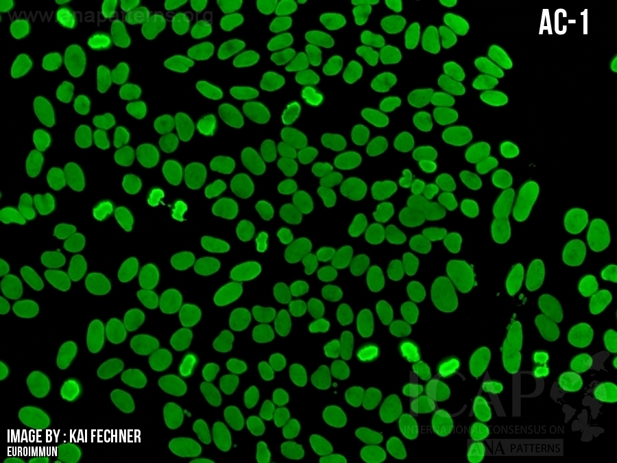
Homogeneous staining of the nucleus with greater intensity of the outer rim and no staining of the metaphase and anaphase chromatin. Accentuation of fluorescence where adjacent cells touch each other may be observed. |
| 抗原相关性 | 板层素A,B,C,板层素相关蛋白,或板层素 B 受体(LBR)。 | Lamins A,B,C, lamin-associated proteins, or lamin B receptor (LBR). |
-
▶AC‑11 核型在常规自身抗体检测中较少见,可见于自身免疫性血细胞减少症、自身免疫性肝病、带状硬皮病、抗磷脂综合征、系统性自身免疫性风湿疾病,也可出现在无明显自身免疫病的个体;目前关于临床相关性的资料主要基于病例报告和小样本队列研究 [1‑5]。▶识别的抗原包括板层素 (A、B、C) 和 LAP‑2;目前尚无针对这些自身抗体的特异性免疫检测商品化试剂盒 [1‑4]。▶The AC-11 pattern is infrequently found in routine autoantibody testing and has been described in autoimmune-cytopenia, autoimmune liver diseases, linear scleroderma, anti-phospholipid syndrome, systemic autoimmune rheumatic diseases, and in individuals with no apparent autoimmune disease; current information on clinical associations is based mainly on case reports and small cohorts [1-5].▶Antigens recognized include lamins (A, B, C) and LAP-2; specific immunoassays for these autoantibodies are currently not commercially available [1-4].
-
二级信息Second level information
-
▶板层素 B 受体(LBR)抗体可见于原发性胆汁性胆管炎(PBC)[6]。▶LBR antibodies have been described in PBC [6].
-
参考文献
-
2026 Topic Review:Konstantinov KN, Konstantinov NK, de Melo Cruvinel W, Andrade LEC, Klotz W, Fritzler MJ. Autoantibodies to the nuclear envelope: Functional anatomy of the HEp-2 cell substrate. Autoimmun Rev. 2026:104154.1.Coppo P, Clauvel JP, Bengoufa D, Fuentes V, Gouilleux-Gruart V, Courvalin JC, Lassoued K. Autoimmune cytopenias associated with autoantibodies to nuclear envelope polypeptides. Am J Hematol. 2004;77:241-92.Konstantinov K, Foisner R, Byrd D, Liu FT, Tsai WM, Wiik A, Gerace L. Integral membrane proteins associated with the nuclear lamina are novel autoimmune antigens of the nuclear envelope. Clin Immunol Immunopathol. 1995;74:89-993.Reeves WH, Chaudhary N, Salerno A, Blobel G. Lamin B autoantibodies in sera of certain patients with systemic lupus erythematosus. J Exp Med. 1987;165:750-624.Senecal JL, Raymond Y. Autoantibodies to DNA, lamins, and pore complex proteins produce distinct peripheral fluorescent antinuclear antibody patterns on the HEp-2 substrate. Arthritis Rheum. 1991;34:249-515.Zhang W, Zhang C, Chen P, Yang C, Gan X, Hussain M, Xun Y, Tian Y, et al. Circulation autoantibody against Lamin A/C in patients with Sjogren's syndrome. Oncotarget. 2016;7:80252-616.Courvalin JC, Lassoued K, Worman HJ, Blobel G. Identification and characterization of autoantibodies against the nuclear envelope lamin B receptor from patients with primary biliary cirrhosis. J Exp Med. 1990;172:961-7.
FAQ
-
细胞核周阳性但中心阴性?有时可观察到细胞核的外周有类似阳性的染色,而细胞核中心是阴性的,这种现象的原因是什么?
核膜型(AC-11和AC-12)会在分裂间期细胞的细胞核周围边界处呈现独特染色,同时由于分裂期核膜破裂,分裂期细胞中没有染色。有时分裂间期的细胞核上可能有微弱染色,为核膜染色叠加所致。除AC-11和AC-12代表的靶抗原外,尚无其他已知的仅位于HEp-2细胞核外周的自身抗原。偶尔有某些样本会呈现核均质染色(AC-1),在核膜附近略有增强,根据我们的经验,使用其他品牌或同品牌不同批次的HEp-2玻片再次检测不会观察到该现象。因此这种特定的荧光分布可能是由细胞培养及固定条件产生的伪影。另外也可能是在IFA操作的孵育期间,细胞基质意外干燥引起的伪影。日期:2019年04月05日